







Transmembrane Protein Services
Introduction to Transmembrane Proteins
Transmembrane proteins (TPs), also known as integral membrane proteins, account for 70%–80% of total membrane proteins. Their main characteristic is that they are insoluble in water. Their amino acid composition is highly hydrophobic, although they also contain hydrophilic amino acids. The portions composed of hydrophobic amino acids penetrate into the hydrophobic region of the lipid bilayer and covalently bind to fatty acid chains. They can be distributed within the lipid bilayer or span the entire membrane. Transmembrane proteins play important roles in fundamental physiological processes, including molecular transport, signal transduction, and energy utilization. On one hand, they can transmit signals from the external environment to cells. On the other hand, they act as receivers to collect signals about the external environment.
Classification of Transmembrane Proteins
Transmembrane proteins can be classified by function into multiple types, including G protein-coupled receptors (GPCRs), ion channels, transporters, and other types of receptors. These proteins play different roles in cells, such as in signal transduction, substance transport, and cell communication. The extracellular region of GPCRs mediates receptor–ligand interactions; activated receptors transmit signals into the cell and recruit downstream effector proteins. Ion channels form a pore in the cell membrane, with hydrophobic amino acids facing the phospholipid bilayer, allowing ions to pass through this channel and cross the hydrophobic core of the cell membrane. Although these transmembrane proteins have different functions, they each play unique and indispensable roles in organisms.
Transmembrane Proteins as Drug Targets
Transmembrane proteins are currently the most important drug targets, accounting for more than 60% of known drug targets at present, and for antibody drug targets, membrane proteins account for nearly 90% or more. Innovative drug R&D targeting transmembrane proteins has long been a research hotspot in the pharmaceutical field, among which ion channels, transporters, GPCRs, and kinases are the largest class of drug targets in drug development [1,2]. They participate in mediating many important cell biological effects, and their dysfunction is associated with various diseases. Therefore, antibodies developed against these targets have unique advantages in treating and preventing related diseases.
The mechanisms of action of antibodies targeting different types of transmembrane proteins differ [3], as shown in the figure below: (a) Antibodies activate GPCRs by acting as ligands to activate or promote dimerization; antibodies inhibit GPCRs, for example, by competing with ligands for binding sites. (b) Promoting internalization is one way antibodies target ion channels. (c) Specific antibodies targeting particular functional states target the inward- and outward-open states of glucose uptake by cells. (d) The left panel shows the binding site of an antibody against MMP-14, and the right panel shows inhibition of enzyme activity by stabilizing an inactive conformation (such as the CD73 dimer).
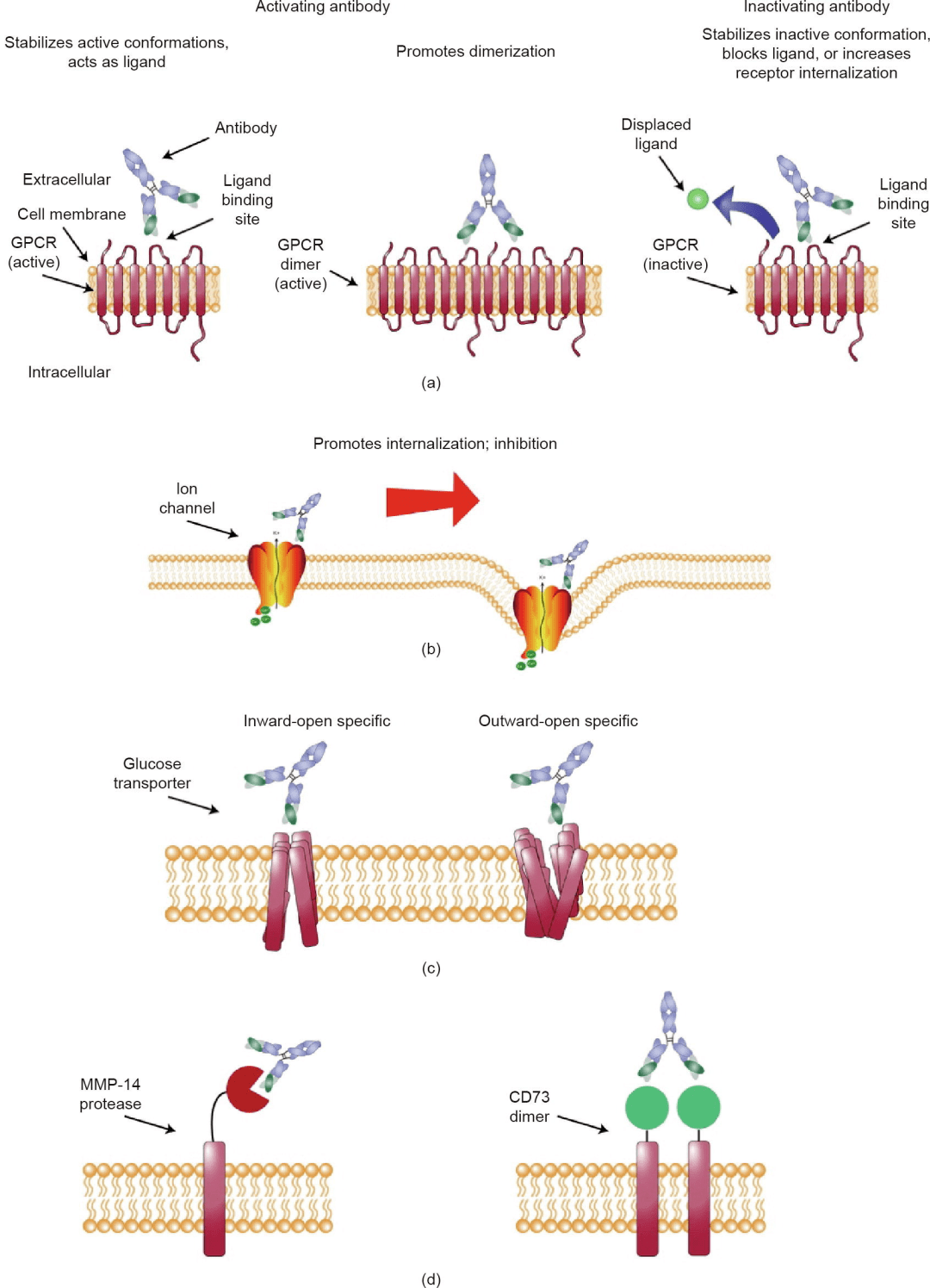
Figure 1: Strategies for selecting antibodies against membrane proteins [3]
Technical Challenges in Developing Multi-pass Transmembrane Proteins
Antigen preparation is the primary requirement for developing antibody drug candidate molecules; an ideal antigen should contain all key sites relevant to the function of the target protein. Therefore, membrane protein antigens need to have the complete conformation and post-transcriptional modifications required for relevant functions; however, the preparation of transmembrane proteins faces many bottlenecks, including low expression levels, poor stability, low solubility, and problems encountered during purification.
• Low Expression Levels:Compared with single-pass transmembrane proteins or secreted proteins, multi-pass transmembrane proteins generally have lower expression levels and need to be embedded in the phospholipid bilayer of the cell membrane to perform their normal functions, whereas overexpressed transmembrane proteins can damage cells.
• Difficult Purification:Multi-pass transmembrane proteins have hydrophobic domains, often have low solubility in solution, and readily aggregate and lose activity, increasing the difficulty of transmembrane protein extraction and purification.
• Complex Structure:Multi-pass transmembrane proteins have complex structures, including multiple transmembrane regions, loop structures, and glycosylation modifications, all of which increase the difficulty of preparing transmembrane proteins.
Jinxuan Biotech Multi-pass Transmembrane Protein Development Platform
1. ECL Technology Platform
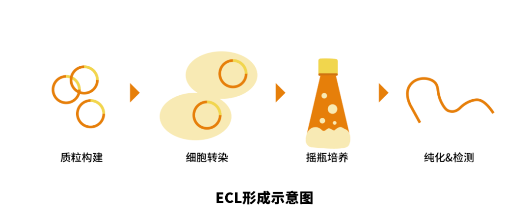
Membrane protein antigens need to have the complete conformation and post-transcriptional modifications required for relevant functions; expressing the extracellular N-terminus or loop domain is simple and relatively high in expression, making it one of the effective methods.
Jinxuan Biotech uses a mammalian cell expression system, which can guide correct protein folding and provide complex N-glycosylation, accurate O-glycosylation, and other post-translational processing functions. As a result, the expressed products are closest to natural higher organism protein molecules in molecular structure, physicochemical properties, and biological function.
Technical Route
Multi-pass transmembrane protein ECL scheme design → plasmid construction and extraction → expression scheme screening → protein purification → activity assay & quality control
2. Detergent Technology Platform
Multi-pass transmembrane proteins cross the cell membrane multiple times and form multiple ECLs (extracellular loops) outside the cell. For example, Claudin18.2 and CD20 both have two ECLs, and each ECL has specific functions and may even interact with each other. Expressing only the ECL makes it difficult to mimic the native conformation of the protein and may cause the loss of some antigen conformational epitopes. For example, the therapeutic CD20 antibodies rituximab and ofatumumab bind different CD20 antigen epitopes. Full-length multi-pass transmembrane proteins can ensure the complete native conformation of the protein, with ECLs fully displayed, making them an excellent choice for screening ideal therapeutic antibodies targeting multi-pass transmembrane protein antigens.
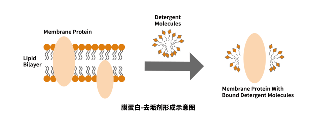
However, transmembrane proteins have hydrophobic domains and bind tightly to lipid membranes, requiring detergents to wash them off the membrane. Detergents have both hydrophilic polar groups and hydrophobic nonpolar groups, providing a hydrophobic environment for membrane proteins in a delipidated state in solution and maintaining the hydrophobic transmembrane structure of membrane proteins.
Based on mammalian cell expression technology, Jinxuan Biotech has established expression and purification platforms for full-length multi-pass transmembrane proteins and has screened several highly effective detergents that can solubilize and purify transmembrane proteins and maintain their native, correct conformation in detergent-containing solutions.
Technical Route
Multi-pass transmembrane full-length protein scheme design → plasmid construction and extraction → expression scheme screening → protein purification → activity assay & quality control
3. VLP Technology Platform
VLP is short for virus-like particle. VLPs are derived from the outer capsid proteins of viruses and are nanoscale particles formed by the automatic assembly of one or more capsid protein components. Because VLP particles are self-assembled from viral capsid proteins, they do not contain a viral genome, and their special spatial structure can induce strong immune responses. These properties make VLPs suitable as recombinant subunit vaccines [4].
VLPs are mainly divided into enveloped VLPs (eVLPs) and non-enveloped VLPs. When VLPs are assembled from the capsid proteins of enveloped viruses, they bud from host cells and become wrapped by the cell membrane, enabling transmembrane proteins on the cell membrane to be displayed on the VLPs. Based on the efficient activation of humoral and cellular immune responses by VLPs, effective antibodies can be obtained even when animals are immunized with antigens/peptides of weak immunogenicity.
Technical Route
Multi-pass transmembrane full-length protein scheme design → plasmid construction and extraction → VLP expression and display → VLP enrichment → activity assay & quality control
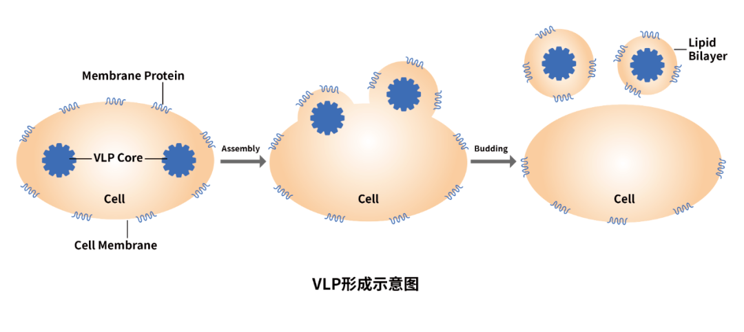
Table 1: Comparison of Advantages and Disadvantages of Different Technology Platforms
For trial use/customization of multi-pass transmembrane protein products, welcome to inquire!
Related Product Introductions
1. Claudin18.2
Claudin18.2 (CLDN18.2) is a member of the Claudin protein family and is located on the cell membrane surface. It consists of two extracellular loops (ECLs), four transmembrane domains, and one cytoplasmic domain. Both the C-terminus and N-terminus of CLDN18 are located in the cytoplasm. Under normal conditions, it is expressed only at low levels in differentiated epithelial cells of the gastric mucosa, but under pathological conditions, Claudin18.2 expression is significantly upregulated in various tumors. In addition, Claudin18.2 is often overexpressed in gastroesophageal cancer, pancreatic cancer, lung cancer, and other solid tumors, making it a popular target with potential for cancer treatment. According to PharmCube database statistics, there are currently about 97 CLDN18.2 drugs in development worldwide, involving different drug types such as monoclonal antibodies, bispecific/multispecific antibodies, cell therapies, and ADCs.
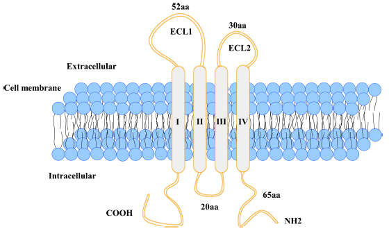
Figure 2: Schematic structure of Claudin18.2 [5]
|
Cat. No. |
Product Name |
Protein Structure |
Technology Platform |
|
C17D |
Recombinant Human CLDN18.2 (N-Twin strep-Flag) |
Full-length |
Detergent Technology |
Partial Data Display

Immobilized Human CLDN18.2-Twin strep-Flag at 5μg/ml (100 μl/well) can bind Anti-Human Claudin18.2 mAb. The ED50 of Anti-Human Claudin18.2 mAb is 29.75 ng/ml.
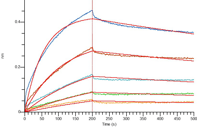
Loaded Anti-Human Claudin18.2 mAb on AHC Biosensor, can bind Human Claudin18.2-Flag with an affinity constant of 6.76 nM as determined in BLI assay.
2. Napi2b
NaPi2b (type IIb sodium-dependent phosphate transporter) is a multi-pass membrane protein and a member of the solute carrier SLC34 family, which consists of NaPi-IIa (SLC34A1), NaPi-IIb (SLC34A2), and NaPi-IIc (SLC34A3). Among them, NaPi-IIb protein mediates inorganic phosphate entry into epithelial cells through sodium ion cotransport and is responsible for maintaining systemic phosphate balance. NaPi2b is widely expressed in ovarian cancer and non-small cell lung adenocarcinoma but is less expressed in normal tissues.
Representative investigational drug: Mersana's upifitamab rilsodotin (XMT-1536, UpRi)
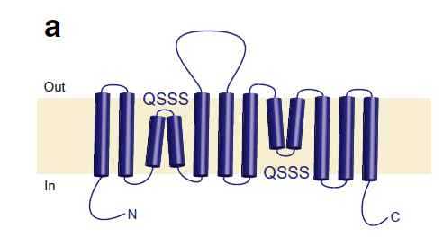
Figure 3: Schematic structure of NaPi2b [6]
|
Catalog Number |
Product Name |
Protein Structure |
Technology Platform |
|
C27G |
Recombinant Human NaPi2b ECL2 (C-Fc) |
ECL2 |
ECL Technology |
|
C27K |
Recombinant Human NaPi2b (C-6His-Flag) |
Full-length |
Detergent Technology |
|
C27W |
Recombinant Mouse NaPi2b (C-Flag) |
Full-length |
Detergent Technology |
|
C27X |
Recombinant Cynomolgus NaPi2b (C-Flag) |
Full-length |
Detergent Technology |
|
Pre-order |
Recombinant Human NaPi2b (VLP) |
VLP |
VLP Technology |
Partial Data Display
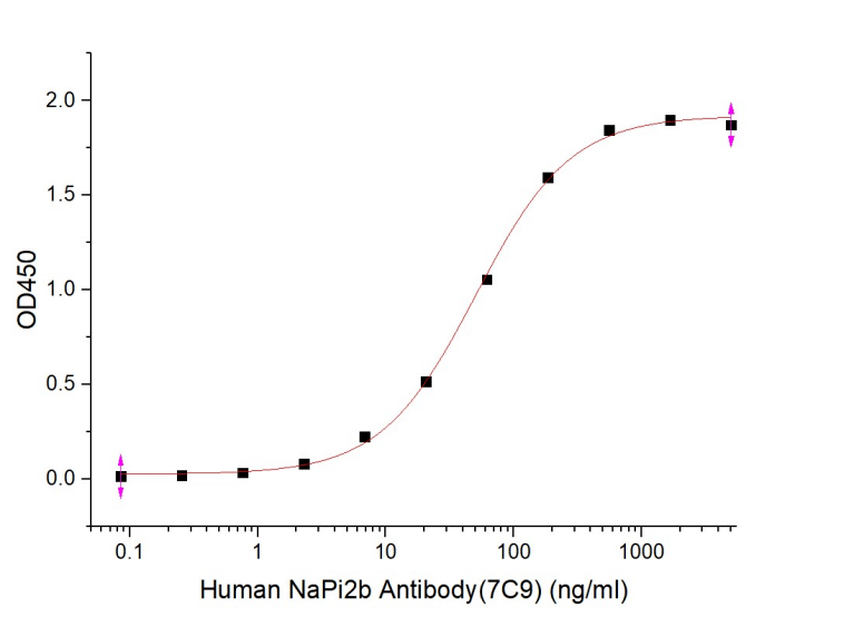
Immobilized Human NaPi2b ECL2-Fc at 2μg/ml (100 μl/well) can bind Human NaPi2b Antibody(7C9) : Biotinylated by NHS-biotin prior to testing. The ED50 of Human NaPi2b Antibody(7C9) is 50.90 ng/ml.
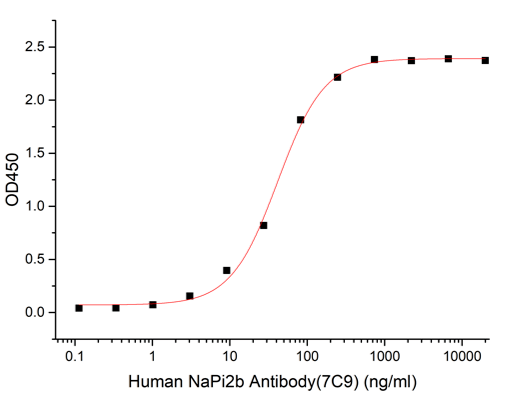
Immobilized Recombinant Human NaPi2b (VLP) at 2μg/ml (100 μl/well) can bind Human NaPi2b Antibody (7C9). The ED50 of Human NaPi2b Antibody (7C9) is 41.35ng/ml.
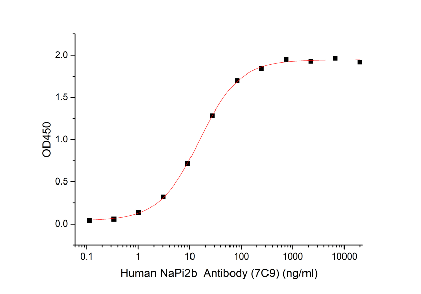
Immobilized Recombinant Human NaPi2b-6His-Flag at 2μg/ml (100 μl/well) can bind Anti-Human NaPi2b Antibody. The ED50 of Anti-Human NaPi2b Antibody is 15.35ng/ml.
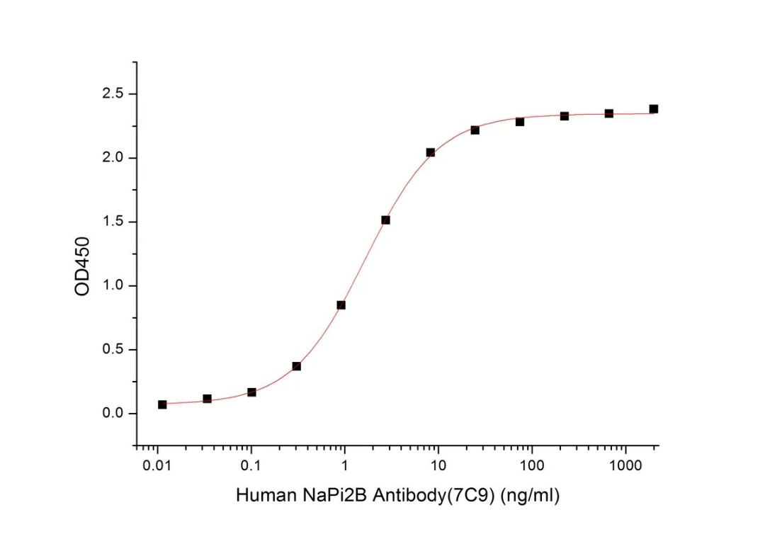
Immobilized Recombinant Mouse NaPi2b-Flag at 2μg/ml (100 μl/well) can bind Human NaPi2b Antibody (7C9). The ED50 of Human NaPi2b Antibody (7C9) is 1.65ng/ml.
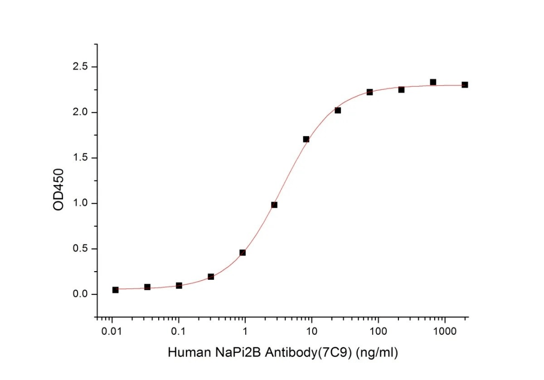
Immobilized Recombinant Cynomolgus NaPi2b-Flag at 2μg/ml (100 μl/well) can bind Human NaPi2b Antibody (7C9). The ED50 of Human NaPi2b Antibody (7C9) is 3.61ng/ml.
3. CD20
CD20 is a non-glycosylated phosphoprotein located on B cells. It belongs to the MS4A protein family (membrane-spanning 4-domains subfamily A) and is mainly expressed from the pre-B cell to mature B cell stage. The CD20 protein consists of four hydrophobic transmembrane domains, one intracellular domain, and two extracellular domains (a large loop and a small loop), with both the N-terminus and C-terminus located in the cytoplasm. CD20 is expressed on the surface of normal and malignant B lymphocytes but is not expressed in hematopoietic stem cells, plasma cells, and other normal tissues. Therefore, CD20 is considered an important target for treating B-cell malignancies and autoimmune diseases. According to PharmCube database statistics, many domestic and international companies are conducting drug R&D targeting CD20, involving different drug types such as monoclonal antibodies, bispecific/multispecific antibodies, cell therapies, and ADCs. More than 150 research pipelines are in preclinical or clinical stages, and more than 50 drugs have been approved for marketing.
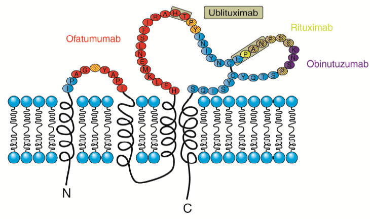
Figure 4: Schematic structure of CD20 [7]
|
Catalog Number |
Product Name |
Protein Structure |
Technology Platform |
|
C17G |
Recombinant Human CD20 (C-Flag) |
Full-length |
Detergent Technology |
|
C20P |
Recombinant Human CD20 (C-6His-Flag) |
Full-length |
Detergent Technology |
|
Pre-order |
Recombinant Human CD20 (VLP) |
VLP |
VLP Technology |
Partial Data Display

Immobilized Human CD20-Flag at 2μg/ml (100 μl/well) can bind Anti-Human CD20 mAb. The ED50 of Anti-Human CD20 mAb is 11.09 ng/ml.

Loaded Anti-Human CD20 mAb on AHC Biosensor, can bind Human CD20-Flag with an affinity constant of 3.47 pM as determined in BLI assay.
References
【3】Georgina To’a Salazar et al. “Antibody Therapies Targeting Complex Membrane Proteins.” Engineering vol. 7,11 (2021) : 1541-1551. doi: 10.1016/j.eng.2020.11.013


