


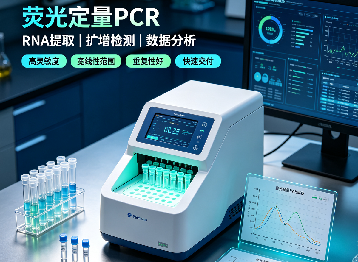




Real-Time Quantitative PCR (qPCR)
In real-time quantitative PCR (qPCR), the most commonly used methods are the non-specific DNA-binding dye SYBR Green I method and the specific TaqMan hydrolysis probe method.
Theoretically, PCR is an exponential amplification process; however, the actual PCR amplification curve is not a standard exponential curve but an S-shaped curve.
This is because as PCR cycles increase, the amplification scale rapidly expands, and various PCR components—such as Taq polymerase, dNTPs, primers, and even the DNA template—gradually become insufficient. PCR efficiency decreases, and the rate of product accumulation gradually slows.Once all Taq polymerase is saturated, PCR enters the plateau phase.Due to the complex interactions of various environmental factors, the timing of entry into the plateau phase and the plateau level vary greatly among different PCR reaction systems, making them difficult to control precisely.Therefore, even in 96 replicate PCR experiments performed under essentially identical conditions, the results can differ considerably. Thus, during an experiment, we cannot directly calculate the initial DNA copy number from the amount of final PCR product.
The fluorescent chemistries used in real-time PCR can be divided into two types: fluorescent probes and fluorescent dyes.Accordingly, real-time PCR methods can be divided into specific and non-specific categories. Specific detection methods use gene-specific oligonucleotide probes labeled with fluorescent dyes in the PCR reaction to detect products, whereas non-specific detection methods add an excess of fluorescent dye to the PCR reaction system; after the dye specifically intercalates into double-stranded DNA, it emits a fluorescent signal.The former has higher specificity due to the additional probe recognition step, while the latter is simpler and easier to perform.
Non-specific SYBR Green I Dye Method
SYBR Green I is a dye with a green excitation wavelength that binds to the minor groove region of all dsDNA double helices. In its free state, SYBR Green I emits weak fluorescence, but once it binds to double-stranded DNA, its fluorescence is greatly enhanced (see mechanism diagram).Therefore, the fluorescence signal intensity of SYBR Green I is correlated with the amount of double-stranded DNA, and the amount of double-stranded DNA present in the PCR system can be detected based on the fluorescence signal.
Specific TaqMan Hydrolysis Probe Method
TaqMan real-time PCR technology is based on TaqMan fluorescent probes. A TaqMan fluorescent probe is an oligonucleotide labeled at both ends with a fluorescent reporter group and a fluorescent quencher group.When the probe is intact, the fluorescence signal emitted by the reporter group is absorbed by the quencher group.
During PCR amplification, the 5′→3′ exonuclease activity of Taq polymerase cleaves and degrades the probe, separating the fluorescent reporter group from the fluorescent quencher group, so that the fluorescence monitoring system can receive the fluorescence signal. That is, each time a DNA strand is amplified, one fluorescent molecule is formed, achieving complete synchronization between the accumulation of fluorescence signal and the formation of PCR product.This enables quantification. Commonly used fluorescent groups are FAM, TET, VIC, and HEX.


