


Apoptosis Detection Service
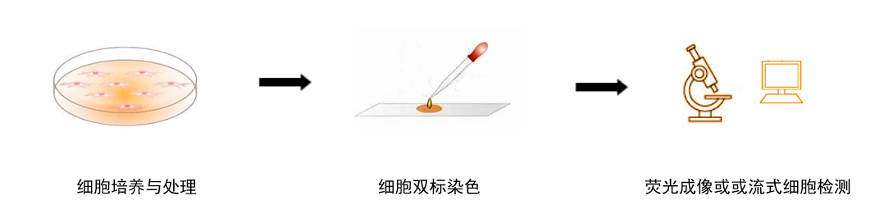
Gxene Biotech has been dedicated to establishing a comprehensive cell culture platform for many years, and we can provide customers with a variety of basic cytology experiments. Relying on superior technical equipment and complete platform construction, we can provide customers with apoptosis detection services. Our scientists have accumulated rich experience in cell technology research. With their skilled and superb technical level, they can quickly complete designated experiments in a very short time, while ensuring fine control of every cell experiment step, ensuring that customers obtain ideal experimental results as much as possible. In addition, our laboratory has advanced immunofluorescence microscopes, flow cytometers, and other systems, which can provide customers with high-quality imaging technology to enhance your research results. According to the actual requirements of customers, Gxene Biotech can establish a complete experimental plan and provide one-stop experimental services from cell processing and slide preparation, fluorescence microscope observation, and photography, improving customer experience.
Apoptosis refers to the autonomous, orderly death of cells controlled by genes to maintain internal environment stability. As a basic biological phenomenon of cells, apoptosis is a death process that the organism actively seeks to better adapt to the living environment. It is a highly orderly, gene-controlled process involving a series of enzymes. It involves the activation, expression, and regulation of a series of genes. The general process includes four stages: induction and initiation, intracellular regulation, execution, and phagocytic clearance of apoptotic cells. The apoptosis detection service provided by Gxene Biotech can be used to detect apoptosis in different cell types and at different stages and to analyze the proportion of apoptotic cells. Detection methods differ depending on the stage of apoptosis.
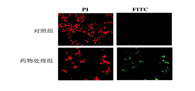
The apoptosis detection provided by Gxene Biotech is mainly based on the "PI and Annexin-V double staining" method. Annexin-V is a Ca2+-dependent phospholipid-binding protein with a molecular weight of 35–36 KD, which can specifically bind to PS with high affinity. We label Annexin V with fluorescein and use the labeled Annexin V as a fluorescent probe. Fluorescence microscopy or flow cytometry can be used to detect the occurrence of apoptosis. Propidium iodide (PI) is a nucleic acid dye that cannot penetrate intact cell membranes, but for cells in the middle and late stages of apoptosis and dead cells, PI can penetrate the cell membrane and stain the nucleus red. Therefore, by using Annexin V-PI double staining technology, cells at different stages of apoptosis can be distinguished. At the same time, relying on its own immunofluorescence imaging platform, Gxene Biotech can provide customers with high-quality image data.
Apoptosis Detection Service — Annexin V /PI Double Staining Method:
|
Item |
Apoptosis Detection Service |
|
Experimental Principle |
In the early stage of apoptosis, phosphatidylserine(Phosphatidyl serine, PS)located on the inner side of the cell membrane migrates to the outer side of the cell membrane. Phospholipid-binding proteinV(Annexin V) is a calcium-dependent phospholipid-binding protein that has high binding affinity forPS. Therefore,Annexin Vcan be used as a probe to detect phosphatidylserine exposed on the outer side of the cell membrane. Thus, usingAnnexin Vwith high affinity forPS,Annexin Vis labeled with fluorescein (such as fluorescein isothiocyanateFITC), and apoptotic cells can be detected by flow cytometry. ThePIdye can bind to intracellularDNA, distinguishing necrotic cells from live cells. The cell membranes of early apoptotic cells and live cells remain intact, and PIdye cannot freely pass through the cell membrane into the cell to bind toDNA, soPIdye cannot label apoptotic cells and live cells. However,PIdye can pass through the cell membrane of necrotic cells and bind to intracellularDNA. AfterPIdye in necrotic cells is excited by a488nmlaser, it emits red fluorescence, which is received by the corresponding channel. Therefore, usingAnnexin VandPItogether can distinguish live cells, early apoptotic cells, and necrotic cells. |
|
Advantages |
·More sensitive and practical for detecting early apoptosis ·Annexin V /PIdouble staining method is more time-saving and simple to perform, and the results are more reliable. It is currently the most ideal method for detecting apoptosis. |
|
Applications |
Apoptosis detection can be applied to: ·Research on different types of pathological specimens, including tumor cells and tissues ·Clinical diagnosis and treatment, new drug development, biological product development, tumor radiotherapy and chemotherapy, and exploration of tumor gene therapy from the perspective of pro-apoptosis ·Early detection of related diseases and evaluation of the efficacy of radiotherapy and chemotherapy |
Main Experimental Workflow of Apoptosis Detection Service:

Our Service Advantages:
–Ensure cell viability and produce high-quality cell climbing slides;
–Comprehensive experimental operation level and experimental workflow; the experimental personnel have many years of successful experimental experience and rich knowledge reserves in cell research, ensuring objective and accurate detection results;
–Provide detailed and complete experimental reports, including original experimental results and each experimental parameter, which can be directly used for paper writing;
–Complete instrument platform: equipped with multiple types of microscopes, flow cytometers, and other important instruments, featuring high resolution, etc., saving precious samples while obtaining detailed data;
–Provide one-stop technical services such as cell culture, cell staining, and photo taking
Experimental Cases: